Investigator: M. Hain
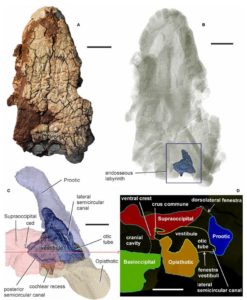
High-resolution X-ray microtomography methods have helped evolutionary biology (a team led by Prof. Klembara) to solve the problem existing over a century, related to the origin of amniotes and thus an essential stage in vertebrate evolution. This stage of evolution is important because the tetrapods broke their ties with the aquatic environment and began to settle the land. Two groups of early tetrapods and ancestors of amniotes – the fossil findings of representatives of diadectomorphs and seymouriamorphs – were analysed by X-ray microtomography, namely their labyrinth of the inner ear. Acquired 3D data from microCT measurements were then subjected to mathematical processing and time-consuming data segmentation with the goal to distinguish fossil structures from the sediment. The results were finally subjected to a cladistic analysis and provided paleobiologists with the knowledge to determine the new classification of these groups in the developmental cladogram.
Result applicator:
Prírodovedecká fakulta UK

Fig.: Fossilised skull of Diadectes absitus and microtomographic virtual 3D reconstruction of inner ear structures
Publications:
- KLEMBARA, J. – HAIN, Miroslav – RUTA, M. – BERMAN, D.S. – PIERCE, S.E. – HENRICI, A.C. Inner ear morphology of diadectomorphs and seymouriamorphs (Tetrapoda) uncovered by high‐resolution x‐ray microcomputed tomography, and the origin of the amniote crown group. In Palaeontology, 2020, p. 1-24. ISSN 0031-0239. (2.632-IF2018) Q1 https://onlinelibrary.wiley.com/doi/full/10.1111/pala.12448
- KLEMBARA, J. – HAIN, Miroslav – ČERŇANSKÝ, A. The first record of anguine lizards (Anguimorpha, Anguidae) from the early Miocene locality Ulm – Westtangente in Germany. In Historical Biology, 2019, vol. 31, no. 8, p. 1016-1027. ISSN 0891-2963. (1.489-IF2018) Q2, https://doi.org/10.1080/08912963.2017.1416469
- ČERŇANSKÝ, A. – YARYHIN, O. – CICEKOVÁ, J. – WERNEBURG, I. – HAIN, Miroslav – KLEMBARA, J. Vertebral comparative anatomy and morphological differences in anguine lizards with a special reference to Pseudopus apodus. In The Anatomical Record, 2019, vol. 302, no. 2, p. 232-257. ISSN 1932-8486. (1.329-IF2018) Q2, https://doi.org/10.1002/ar.23944
 Contacts
 Intranet
 SK
SK